
Microscopy
Already have a Logon Account?
|
Welcome
Welcome to the University of Research and Development FBS Portal. This site is designed to automate the use of our Core Facilities and to provide the best possible customer service. Quick Info
For more info, please contact the Priority Software Support Team. Schedules
| Our Core Facilities To learn more about a particular facility or to request access, please click on a facility name below. |
 Resource Calendar
 Live Usage
Main Contact Info
Garth Roberts
Priority Software Inc.
9006 4th Avenue South
Birmingham, AL 35206
Remittance Contact Info
Henry Rhoe
Priority Software Inc.
9006 4th Avenue South
Birmingham, AL 35206
| Other Contacts | |||
|---|---|---|---|
| Caleb Smith | Manager | (205) 202-8400 | CalebSmith@demodata.com |
| Shawn Talbot | Staff | (205) 202-8400 | ShawnTalbot@demodata.com |
This facility has not published any Products. Please check back.
The following Products and Services are available within our facility:
Instruments
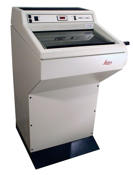 |
CryostatMicrom HM550 Cryostat is used for thick and thin sectioning of cryo-preserved and embedded specimens. The instrument will cut sections of 5 microns up to 500 microns at at temperature as low as -40 Celsius. |
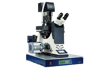 |
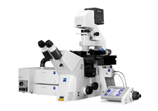
Nikon Fl StereoThe Leica MZ16 Fluorescence Stereomicroscope is designed for acquisition of fluorescent images of many dyes including DAPI, FITC, TRITC, CFP and YFP at long working distance and low magnification (4X - 20X). The instrument has a black and white camera for fluorescent image acquisition and a color camera for imaging of vital dyes. The stage is motorized and is capable of X, Y and Z montage imaging. |
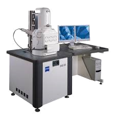 |
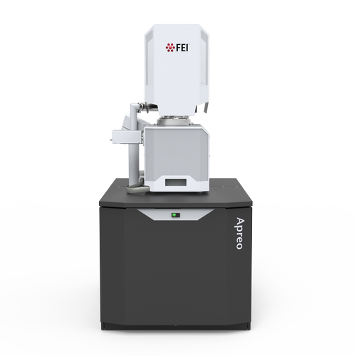
SEMBased on Zeiss' GEMINI field emission SEM column, the Zeiss Supra 40V Scanning Electron Microscope (SEM) provides ultra high resolution at low kV. Additionally, the variable presser chamber allows for imaging of non-conductive samples. The large chamber and user friendly software make this a versatile easy to use instrument. |
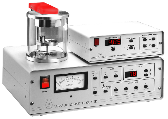 |
Sputter CoaterBal-Tec MED 20 High Vacuum Coater - This quick turbo-pumped unit with accessory for rotary and directional electron beam evaporation. Useful for low-angle high-resolution tungsten shadowing. |
 |
Super Resolution ConfocalThe Zeiss LSM 710/Elyra S.1 is a single microscope stand that functions as both a laser scanning confocal and a structured illumination super resolution microscope. |
 |
TEMThe FEI Tecnai Transmission Electron Microscope (TEM) is a 20-120kV instrument that we typically operate at 80kV for improved contrast of biological specimens. The BioTwin lens configuration enhances specimen contrast at the medium magnification used with resin-embedded tissue while permitting sub-nanometer resolution with the right specimen and instrument configuration. |
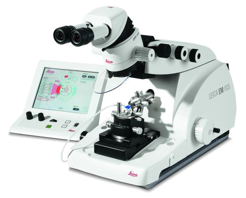 |
UltramicrotomeThe Leica UCT is used for thick and thin sectioning of resin-embedded specimens. With good lighting, precise orientation of specimen and knife, plus the control of approach and feed from a separate unit, the UCT will cut sections as thin as 50nm for TEM or quickly cut 1-5 micron sections for light microscopy. Nearby in the lab is a knife breaker for making glass knives. We also have a cryo-chamber for sectioning at temperatures below -100C. |
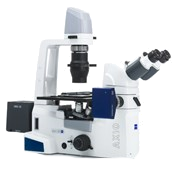 |
Zeiss FLMThe Zeiss Axiovert 200M is a widefield fluorescent microscope fitted with a 100W Hg lamp variable intensity power supply. The instrument has fluorescence filter cubes for a wide range of dyes including DAPI, CFP, FITC, GFP, YFP, TRITC, CY5 and CFP/YFP FRET. Brightfield and DIC imaging for high magnification is also available. |
Software
 |
Imaris 4-D SoftwareImaris is used for data management, visualization, analysis, segmentation and interpretation of 3D and 4D microscopy datasets. |
This facility has not published any News. Please check back.
Quick Quotes have not been configured. Please check back soon (Code 001, Code 002)
, please enter that email address here.")